At Spine & Neuro Center we provide a ray of hope to people who have none and thereby increase the quality of life. Spine and Neuro Institute is an institution which not just wants to give treatment to the patients but also take into consideration the overall well being of the patient
Spine & Neuro is high qualified Neurosurgical Center that offer comprehensive treatment for common and complex pathology of the brain and spine, with a excellent medical group in a and a warm human touch, with the aim of minimally invasive neurosurgery with the most advanced technology. Diseases resulting from degenerative changes in the nervous system markedly impact the lives of millions and pose growing public health challenges. The prevention and treatment of these neurodegenerative disorders represents critical goals of medical research today and is the mission of Spine and Neuro Center.
Spine & Neuro Center offers the following medical specialties:
Brain treatments: | |
Brain Tumor Resection | |
Deep Brain Stimulation (DBS) procedures for Parkinson disease, dystonias, and other functional Neurosurgeries | |
Neuroendoscopy for intraventricular procedures (hidrocephalus, intraventricular tumors) and Neuroendoscopy assisted Surgeries | |
Stereotactic biopsy and Stereotactic guided surgery | |
Surgery for Epilepsy | |
Trigeminal nerve Decompression (Microvascular decompression or MVD) | |
Neuronavegation biopsy (NON-head pint, same day in and out procedure) and Neuronavegation guided surgery | |
Cervical Spinal treatments: | |
Anterior Cervical Corpectomy and Fusion | |
Cervical anterior fusion or disc replacement | |
Cervical posterior fusion and tumor resection | |
Craneoplasty | |
Endoscopic Pituitary tumor Resection | |
Laminaplasty | |
Microlaminectomy | |
Posterior Cervical Fusion (PCF) | |
Thoracic posterior fusion and transthoracic fusion | |
In the lumbar spine we can offer standard and minimally invasive techniques for: | |
Ballon Vetebroplasty (kyphoplasty) | |
Herniated disc, laminectomy, spinal stenosis without fusion and spinal fusions | |
Interspinous Process Spacers with open or percutaneous technique (minimally invasive technique) | |
Minimally Invasive Spinal Fusion (MIS PLF) | |
Microdiscectomy | |
Posterior Lumbar Interbody Fusion (PLIF) |
An interdisciplinary group integrated by board-certified doctors with different specialties, highly trained nursery personnel will support the diagnosis and treatment of our patients with the most advanced technological equipment.
Your Doctor will recommend a recovery time in the hospital or give you further instructions on how to go through the next days in your hotel and when to come back for further checks.
Your Medical Concierge will get all needed pharmacy shopping done. He will help you through the recovery process.
The only thing you have to do is to get better.
Stereotaxy and neuronavegation are together also called image-guided surgery (IGS), computer-assisted/computer-aided surgery (CAS).
Stereotactic neurosurgery FRAMELESS Stereotaxy involves mapping the brain in a three dimensional coordinate system (3D). With the help of MRI and CT scans and 3D computer workstations, neurosurgeons are able to accurately target any area of the brain in stereotactic space. Stereotactic brain biopsy is a minimally invasive procedure that uses this technology to obtain samples of brain tissue for diagnostic purposes when there are suspicious for tumors or infections.
The main indications for stereotactic biopsy are lesions deep inside the brain, multiple lesions, or lesions in a surgically poor candidate who cannot tolerate general anesthesia.
On the morning of surgery a headring (stereotactic frame) is placed on the patients head with local anesthesia that numbs the skin in four points (2 in the forehead and 2 in the back of the head) and placing the ring on the head with four pins.
A CT scan and or a MRI is then performed.
The stereotactic plan is done with very sophisticated software (Praesis Plus) that gives the surgeon the coordinated of the lesion aimed.
In the operating room, the patient receives light sedation. An incision only 1cm long is made in the scalp and a small hole is drilled into the skull.
A thin biopsy needle is inserted into the brain using the coordinates obtained by the computer workstation.
The specimen is then sent to the pathologist for evaluation. Patients are monitored for several hours following the procedure and usually go home the same day. The risks associated with stereotactic brain biopsy are minimal (intracranial hemorrhage, seizure, or infection). Sometimes the sample of tissue obtained may be non-diagnostic, which may warrant a repeat biopsy.
NeuroNavegation:
Neuronavegation is also called FRAMELESS Stereotaxy because it is similar to the Stereotactic surgery BUT WITHOUT the head frame. The surgical indications are mainly the same as stereotactic surgery BUT this technique has less accuracy (1-2mm) than FRAME BASE Stereotaxy (0.3 to 0.5mm "submilimetric precision").
Using a technology similar to a global positioning system (GPS), neuronavigation provides the surgeon the ability to see a patient’s anatomy in three dimensions and accurately pinpoint a location in the brain or spinal cord with the aid of diagnostic images such as computed tomography (CT) and magnetic resonance (MR). This allows the surgeon to determine lesion borders, delicate structures to reduce the risk of damage.
The term "neuronavigation" is synonymous with image-guided surgery (IGS), computer-assisted/computer-aided surgery (CAS).
As the surgeon moves an instrument in the brain, its position is precisely calculated, that data is then transferred to a computer in the operating room thru camera that is place in the OR. The computer then superimposes the position of the instruments as they are used in surgery onto images of the anatomy displayed on a monitor (CT or MRI), allowing the surgeon to see the exact placement and direction the instrument is moving.
On the morning of surgery a headring (stereotactic frame) is placed on the patients head with local anesthesia that numbs the skin in four points (2 in the forehead and 2 in the back of the neck)
FAQS:
How does neuronavigation technology work?
You might compare surgical neuronavigation technology to the location and directional tracking systems used for cars and ships today -- it is, in effect, a GPS system for the surgeon. Much as the driver of a car uses the GPS system to find the way on the road, the surgeon depends on these images to confirm the position of his or her instruments in the patient's body.
Why is it beneficial for surgeons to use neuronavigation during surgery?
Ultimately, using neuronavigation helps the surgeon accurately detect where he or she is working in the patient's body at every moment during surgery.
This capability enables the surgeon to make smaller incisions. When trauma to the body is minimized, the patient may spend less time in recovery and may experience fewer complications.
How do patients benefit from neuronavigation in brain surgery?
Neuronavigation gives surgeons image-guided precision for delicate procedures like tumor removal or Deep Brain Stimulation (DBS). During tumor removal, navigation and intra-operative imaging allow the surgeon to "see" whether he or she has successfully removed the entire tumor and avoid damage to surrounding healthy tissue. During DBS, the surgeon is able to confidently and precisely target the exact point on the brain necessary for the treatment of Parkinson‚ disease or other neurological disorders.
Artificial disc replacement (ADR) is newer type of spinal disc procedure that utilizes an anterior (front - through the abdominal region) approach to replace a painful, arthritic, worn-out intervertebral disc of the lumbar spine with a metal and plastic prosthesis (artificial disc). Currently, the artificial disc replacement is only FDA-approved in the United States for use in the lumbar (low back) region, although many products are currently being studied for use in the cervical (neck) region.
FAQS:
What is an artificial disc?
The disc is the soft cushioning structure located between the individual bones of the spine, called "vertebra." It is made of cartilage-like tissue and consists of an outer portion, called the annulus, and an inner portion, called the nucleus (Figure 1). In most cases, the disc is flexible enough to allow the spine to bend.
Who needs an artificial disc?
The indications for disc replacement may vary for each type of implant. Some general indications are pain arising from the disc that has not been adequately reduced with non-operative care such as medication, injections, chiropractic care and / or physical therapy. Typically, you will have had an MRI that shows disc degeneration. Often discography is performed to verify which disc(s), if any, is related to your pain. (Discography is a procedure in which dye is injected into the disc and X-rays and a CT scan are taken. See the NASS Patient Education brochure on Discography for more information.) The surgeon will correlate the results of these tests with findings from your history and physical examination to help determine the source of your pain.
Artificial Disc Replacement: Surgery Risks
All surgery carries risks from anesthesia, blood clots and infections. If complications from these risks arise, they most often can be successfully treated. The physical condition of the patient (such as obesity and diabetes) can also add risk to surgery.
Anterior lumbar interbody fusion (ALIF) is a type of spinal fusion that utilizes an anterior (front - through the abdominal region) approach to fuse (mend) the lumbar spine bones together. Interbody fusion means the intervertebral disc is removed and replaced with a bone (or metal) spacer, in this case using an anterior approach. Although the anterior lumbar ALIF approach involves retracting (moving out of the way, temporarily) large blood vessels (aorta, vena cava) and the intestines, there is a wide exposure of the intervertebral disc without retraction of the spinal nerves and neurologic structures.
Post-Operative Care & Recovery Time:
After surgery, the patient will normally stay in the hospital between 2 to 5 days. The specific time of stay in the hospital will depend on the patient and the surgeon's specific post-operative treatment plan. The patient will normally be up and walking in the hospital by the end of the first day after the surgery. The surgeon will have a specific post-operative recovery / exercise plan to help the patient return to normal life as soon as possible.
FAQS:
What is an Anterior Lumbar Interbody Fusion (ALIF)?
Is a surgery to correct problems with your spine (backbone). The spine is made up of bones called vertebrae that are like blocks placed on top of each other. Usually, the spine should be straight when seen from the back and like a letter 'S' from the side.
Why is it done?
Patients who are suffering from chronic back pain due to certain spinal conditions such as degenerative disc disease are potential candidates for the ALIF procedure. Surgery is typically considered only after other non-operative therapies have failed.
What happened after the surgery?
After surgery, the patient will normally stay in the hospital between 2 to 5 days. The specific time of stay in the hospital will depend on the patient and the surgeon's specific post-operative treatment plan. The patient will normally be up and walking in the hospital by the end of the first day after the surgery. The surgeon will have a specific post-operative recovery / exercise plan to help the patient return to normal life as soon as possible.
Anterior lumbar corpectomy and fusion is a special type of spinal decompression and fusion procedure that utilizes an anterolateral (flank or side of the body incision - through the abdominal region) approach to remove bone and tissue that is causing compression of the spinal cord and nerves. This procedure involves removing the entire vertebral body, superior and inferior disc, which must be replaced with a piece of bone graft or metal and mended (fused) together to maintain stability. A small metal plate with screws is used to add additional stability and prevent the graft from moving.
Post-Operative Care & Recovery Time: Most patients are usually able to go home 4-7 days after surgery. Patients will typically stay longer, approximately 6-10 days, if a posterior spinal surgery is also performed. Before patients go home, physical therapists and occupational therapists work with patients and instruct them on proper techniques of getting in and out of bed and walking independently. Patients are instructed to avoid bending at the waist, lifting (more than five pounds), and twisting in the early postoperative period (first 2-4 weeks) to avoid a strain injury. Patients can gradually begin to bend, twist, and lift after 4-6 weeks as the pain subsides and the back muscles get stronger.
FAQS:
What is an anterior lumbar corpectomy and fusion?
Anterior lumbar corpectomy and fusion is a special type of spinal decompression and fusion procedure that utilizes an anterolateral (flank or side of the body incision - through the abdominal region) approach to remove bone and tissue that is causing compression of the spinal cord and nerves. However, in order to do so generally involves removing nearly the entire vertebral body and disc, which must be replaced with a piece of bone graft and mended (fused) together to maintain stability.
What happened after the surgery?
Most patients are usually able to go home 4-7 days after surgery. Patients will typically stay longer, approximately 6-10 days, if a posterior spinal surgery is also performed. Before patients go home, physical therapists and occupational therapists work with patients and instruct them on proper techniques of getting in and out of bed and walking independently. Patients are instructed to avoid bending at the waist, lifting (more than five pounds), and twisting in the early postoperative period (first 2-4 weeks) to avoid a strain injury. Patients can gradually begin to bend, twist, and lift after 4-6 weeks as the pain subsides and the back muscles get stronger.
What are the results?
The results of anterior thoracic/lumbar corpectomy and fusion surgery are generally good and successful. However, the risks are higher than many other types of spinal surgery because patients who require this type of surgery often have a severe spinal condition (tumor, infection, etc.). In addition, patients are frequently older and have other significant medical problems. A number of research studies in medical journals demonstrate greater than 70% good or excellent results from anterior thoracic/lumbar corpectomy and fusion surgery for various spinal conditions.
Anterior cervical discectomy and fusion (ACDF) is performed for patients with a symptomatic, painful herniated disc in the neck. Anterior cervical discectomy and fusion is the most common neck surgery performed by spine surgeons. It is performed to remove the intervertebral disc and the protruding portion that is compressing the spinal cord and / or nerve root. The disc must be replaced with a piece of bone graft, metal, and a plate with screws to maintain stability and prevent the graft from moving.
Surgical Technique: The surgery is performed utilizing general anesthesia. Patients are positioned in the supine (lying on the back) position. A 2-4 centimeter (depending on the number of levels) transverse incision is made in one of the creases of neck, just off the midline. The cervical fascia is gently divided in a natural plane, between the esophagus and carotid sheath (area containing the blood vessels in the neck). Small retractors is placed, some surgeons use microscope and some not. A clear view of the anterior (front part) vertebral body and discs. After the retractor is in place, an x-ray is used to confirm that the appropriate spinal level(s) is identified. A complete discectomy (removal of the disc, including the protruding fragment) is typically performed.
A graft size is chosen so as to restore the normal disc space height and the graft is then gently tapped into the disc space, in between the two vertebral bodies. A small titanium metal plate is frequently placed, affixed to the vertebrae with small screws, to impart immediate stability to the construct and allow for optimal bone healing and fusion. X-rays are then used to confirm appropriate position and alignment of the graft, plate and screws. The deep facial layer and subcutaneous layers are closed with a few strong sutures. The skin can be closed using special surgical glue, or a few invisible stitches. A small bandage is recommended for a few days to keep the wound clean. The total surgery time is approximately 2 to 3 hours, depending on the number of spinal levels involved.
Post-Operative Care & Recovery Time: Most patients are able to go home 1-2 days after surgery. Patients are instructed to avoid bending and twisting of the neck in the early postoperative period (first 2-4 weeks). Patients can gradually begin to bend and twist their neck after 2-4 weeks as the pain subsides and the neck and back muscles get stronger. Patients are also instructed to avoid heavy lifting in the early postoperative period (first 2-4 weeks). Patients can use a cervical soft collar or plastic neck brace depending on the procedure done. This reduces the stress on the neck area and helps decrease pain. The wound area can be washed with water and soap during shower and then left clean and dry, cover with bandage if necessary. Patients should not take a bath until the wound has completely healed, which is usually around 2 weeks after surgery. Patients may begin driving when the pain has decreased to a mild level and mobility of the neck has improved, which is usually between 10-14 days after surgery. Patients need to be able to turn their neck and body enough to see right and left while driving.
Patients may return to light work duties as early as 2-3 weeks after surgery, depending on when the surgical pain has subsided. Patients may return to heavy work and sports as early as 8-12 weeks after surgery, when the surgical pain has subsided and the neck and back strength has returned appropriately. Patients will return for a follow-up visit to see the doctor approximately 8 days after surgery. The incision will be inspected and suture will be removed. Patients will return to see the doctor 4-6 weeks thereafter, and an x-ray will be taken to confirm the fusion area is stable and healing appropriately.
FAQS:
What is an Anterior Cervical Discectomy & Fusion (ACDF)?
Anterior cervical discectomy and fusion (ACDF) is a procedure used to treat neck problems such as cervical radiculopathy, disc herniations, fractures, and spinal instability. In this procedure, the surgeon enters the neck from the front (the anterior region) and removes a spinal disc (discectomy). The vertebrae above and below the disc are then held in place with bone graft and sometimes metal hardware. The goal is to help the bones to grow together into one solid bone. This is known as fusion. The medical term for fusion is arthrodesis.
What do surgeons hope to achieve?
In most cases, ACDF is used to stop symptoms from cervical disc disease. Discs start to degenerate as a natural part of aging and also from stress and strain in the structures of the neck. Over time, the disc begins to collapse, and the space decreases between the vertebrae.
What are the results?
Anterior cervical discectomy is successful in relieving arm pain in 92 to 100% of patients. However, arm weakness and numbness may persist for weeks to months. Neck pain is relieved in 73 to 83% of patients. In general, people with arm pain benefit more from ACDF than those with neck pain. Aim to keep a positive attitude and diligently perform your physical therapy exercises. The results of anterior cervical discectomy and fusion surgery in the treatment of a painful, cervical herniated disc are generally very good. Numerous research studies in medical journals demonstrate greater than 85-95% good or excellent results from anterior cervical discectomy and fusion surgery. Most patients are noted to have quick improvement of their pain and return to normal function following surgery. The most important fact for a good outcome is the time from the beginning of the symptoms to the surgery, patients with long history of neck and arm pain have nerve root damage by disc compression and surgical results are not as good.
Anterior cervical corpectomy and fusion (ACCF) is performed for patients with symptomatic, progressive cervical spinal stenosis and myelopathy. It is performed to remove the large, arthritic osteophytes (bone spurs) that are compressing the spinal cord and spinal nerves. However, in order to do so generally involves removing nearly the entire vertebral body and disc, which must be replaced with a piece of bone graft or metal and mended (fused) together to maintain stability.
Surgical Technique: The surgery is performed utilizing general anesthesia. Patients are positioned in the spine (lying on the back) position, generally using a standard flat operating table. A 2-4 centimeter (depending on the number of levels) transverse incision is made in one of the creases of neck, just off the midline. The cervical fascia is gently divided in a natural plane, between the esophagus and carotid sheath (area containing the blood vessels in the neck). Small retractors and an operating microscope are used to allow the surgeon to visualize the anterior (front part) vertebral body and discs. After the retractor is in place, an x-ray is used to confirm that the appropriate spinal level(s) is identified. A complete corpectomy and discectomy (removal of the cervical vertebral body and disc, including the protruding osteophytes and disc fragments) is typically performed, allowing the spinal cord and nerves to return to their normal size and shape when the compressive lesions are removed. A graft size is chosen so as to restore the normal disc space height and the graft is then gently tapped into the disc space, in between the two vertebral bodies. A small titanium metal plate is frequently placed, affixed to the vertebrae with small screws, to impart immediate stability to the construct and allow for optimal bone healing and fusion. X-rays are then used to confirm appropriate position and alignment of the graft and hardware. The deep facial layer and subcutaneous layers are closed with a few strong sutures. The skin can usually be closed using special surgical glue, or nylon suture and covered with a bandage. The total surgery time is approximately 2 to 3 hours, depending on the number of spinal levels involved.
These procedures are performed at Spine & Neuro qualified Neurosurgical Center a private institution dedicated to the treatments for common and complex pathology of brain and spine. The Neurosurgical Center has the most modern infrastructure, supported by the Galenia hospital in the respective headquarter.
Post-Operative Care & Recovery time:
Most patients are able to go home 1-2 days after surgery. Patients are instructed to avoid bending and twisting of the neck in the early postoperative period (first 2-4 weeks). Patients can gradually begin to bend and twist their neck after 2-4 weeks as the pain subsides and the neck and back muscles get stronger. Patients are also instructed to avoid heavy lifting in the early postoperative period (first 2-4 weeks). Patients can use a cervical soft collar or plastic neck brace depending on the procedure done. This reduces the stress on the neck area and helps decrease pain.
The wound area can be washed with water and soap during shower and then left clean and dry, cover with bandage if necessary. Patients should not take a bath until the wound has completely healed, which is usually around 2 weeks after surgery. Patients may begin driving when the pain has decreased to a mild level and mobility of the neck has improved, which is usually between 10-14 days after surgery. Patients need to be able to turn their neck and body enough to see right and left while driving. Patients may return to light work duties as early as 3-4 weeks after surgery, depending on when the surgical pain has subsided. Patients may return to heavy work and sports as early as 8-12 weeks after surgery, when the surgical pain has subsided and the neck and back strength has returned appropriately. Patients will return for a follow-up visit to see the doctor approximately 8 days after surgery. The incision will be inspected and suture will be removed. Patients will return to see the doctor 4-6 weeks thereafter, and an x-ray will be taken to confirm the fusion area is stable and healing appropriately.
The results of anterior cervical corpectomy and fusion surgery in the treatment of symptomatic, progressive, cervical spinal stenosis and myelopathy are generally good. The surgery serves to improve pain and function and prevent further neurologic deterioration and paralysis. Numerous research studies in medical journals demonstrate greater than 80-90% good or excellent results from anterior cervical corpectomy and fusion surgery. The most important fact for a good outcome is the time from the beginning of the symptoms to the surgery, patients with long history of neck and arm pain have nerve root damage by disc compression and surgical results are not as good.
FAQS:
What is an anterior cervical corpectomy (ACCF)?
Is similar in approach to an ACD, but the procedure itself is more extensive. The USC Center for Spinal Surgery reports that an ACCF is performed to remove larger bone spurs, and may require the removal of an entire vertebrae and / or intervertebral disc. After removal, that portion of the spine is fused together for stability with bone, grafting materials or metal plates.
What are the results of this treatment?
The results of anterior cervical corpectomy and fusion surgery in the treatment of symptomatic, progressive, cervical spinal stenosis and myelopathy are generally good. The surgery serves to improve pain and function and prevent further neurologic deterioration and paralysis. Numerous research studies in medical journals demonstrate greater than 80-91% good or excellent results from anterior cervical corpectomy and fusion surgery. The fusion rate is significantly improved with the use of a small titanium plate, and typically obviates the need for a halo postoperatively. Most patients are noted to have gradual improvement of their pain and function following surgery.
What are the risks and complications?
Technically, a corpectomy is a more difficult spine surgery to perform. Similar to a discectomy, the risks and possible complications of this surgery for cervical spinal stenosis include:
Nerve root damage, Damage to the spinal cord, Bleeding, Infection, Graft dislodgment, Damage to the trachea/esophagus, Continued pain.
However, a corpectomy is a more extensive procedure than a discectomy, so the risks are statistically greater, especially with respect to neurologic issues, bone grafting and bleeding.
Brain tumors can be first biopsy for cell recognition and then be schedule for medical treatment of surgical approach. Other cases when the tumor is well known in the radiographic study, it may go straight in to surgery.
Open brain surgery is widely in all neurosurgical centers for removing brain tumors and it can be assisted by neuronavegation to increase the tumor resection and decrees the risk of damaging important structures.
It can involve a multidisciplinary medical team to achieve the best result for the patient.
FAQS:
What is a brain tumor?
A brain tumor is an abnormal mass of tissue in which some cells grow and multiply uncontrollably, apparently unregulated by the mechanisms that control normal cells. The growth of a tumor takes up space within the skull and interferes with normal brain activity. A tumor can cause damage by increasing pressure in the brain, by shifting the brain or pushing against the skull, and by invading and damaging nerves and healthy brain tissue. The location of a brain tumor influences the type of symptoms that occur. This is because different functions are controlled by different parts of the brain.
What are the common symptoms of brain tumors?
The most common symptoms include headaches, which can be most severe in the morning; nausea or vomiting, which can be most severe in the morning; seizures or convulsions; difficulty thinking, speaking, or finding words; personality changes; weakness or paralysis in one part or one side of the body; loss of balance; vision changes; confusion and disorientation; and memory loss. Different parts of the brain control different functions, so symptoms will vary depending on the tumor's location.
How is a brain tumor identified?
A brain tumor diagnosis usually involves several steps, which can include a neurological examination, brain scan(s) and / or a biopsy.
The Craneoplasty is a surgery done for cosmetic reasons and for brain protection proposes. It involves the neurosurgery repair of irregularity of the skull or a past surgical removal of part of the skull. The surgery involves the use of bone graft from the patient (usually taken off in a previous surgery for many reasons) or a synthetic material such as titanium, carbon, ceramic or polymethyl Methacrylate Implant to repair a gap or defect in the vault of the skull.
Cranioplasty is a procedure that takes approximately two hours to replace a portion of the skull with either original bone tissue or synthetic implants. The procedure is done under general anesthetic and is positioned with the bone defect uppermost. The area of incision is shaved and prepared with antiseptic. The incision is done around the affected area, the scalp is cut apart from the ‘duramater’ (the underlying covering of the brain), and the edges of the surrounding bone are cleaned to let the new graft have good contact with healthy bone.
The patient’s bone graft or a replacement material (that is shaped for the fit gap) is placed in the defect area and secured with screws or plates.
Titanium implant
Patients own bone graft
Carbon Implant
Once the implant is in position and secure, the skin flap is closed either with nylon suture or with staples.
FAQS:
What Is Cranioplasty?
The Cranioplasty is a surgical procedure in which part of the skull is reconstructed. This procedure can be performed for aesthetic or medical reasons, and sometimes a blend of both. It requires a skilled surgeon because there are a number of serious risks.
Why get a Cranioplasty?
One reason to perform a cranioplasty is to address a deformity or defect in the skull. There are a number of conditions which can lead to abnormalities in skull structure, such as cases in which the cranial sutures close early and the skull cannot expand as the brain grows. Likewise, some congenital conditions can cause children to be born with skull anomalies which can be corrected with cranioplasty.
What are the risks of a cranioplasty?
Risks of a cranioplasty can include infection at the surgical site, damage to the nerves in the face, and injuries to the brain. Brain injuries are especially common concerns after trauma, because more of the brain may need to be exposed during surgery and the brain can be subject to swelling which may cause a dangerous rise in pressure inside the skull.
Endoscopic pituitary surgery is performed through a natural nasal air pathway without any incisions (unlike the conventional microscopic surgery performed with an incision made under the upper lip or inner aspect of a nostril). Endoscopic surgery does not require the use of a metallic transsphenoidal retractor that is used for conventional microscopic surgery. A 4-mm endoscope brought in from the nose entry to the base of the sphenoidal sinus, a small window is made with special instruments we have an approach to the base of the skull below the sella turca where the pituitary gland sits.
The tumor is removed with specially designed surgical tools. Postoperative nasal packing is not necessary and postoperative discomfort is minimal. Most patients are able to go home the following day. There are some optical advantages of an endoscope approach such as a wide-angled panoramic view, an angled view by angled lens endoscopes.
FAQS:
How is this surgery performed?
Most pituitary tumors can be removed transsphenoidally. The approach is through the sphenoid sinus, one of the facial air spaces behind the nose. Rarely, a craniotomy is required, where the skull is opened to reach the tumor. There are three basic approaches to the sella, which is the bony cavity in the skull base where the pituitary gland is located. Many neurosurgeons now use a direct transnasal approach, where an incision is made in the back wall of the nose and the sphenoid sinus is entered directly. Finally, it is possible to make an incision under the lip and approach through the upper gum, and enter the nasal cavity and then the sphenoid sinus.
How should I choose a surgeon for my pituitary operation?
It has been shown that the success of surgery is dependent on the amount of experience the surgeon has at performing pituitary operations. Surgeons with the most experience generally have the highest rates of cure, meaning complete tumor removal. In addition, the rate of complications is lowest among experienced pituitary surgeons.
What is the chance of being cured?
It depends upon the type, size and location of the tumor and the expertise of the surgeon. Patients with Cushing's disease usually have small tumors (microadenomas) and are surgically cured about 90% of the time based on data published by expert pituitary surgeons. Patients with acromegaly often have larger, more invasive tumors which are harder to cure surgically. The success rate with growth-hormone secreting macroadenomas is about 60% in the best surgical series.
Laminaplasty is a unique posterior (back of the neck incision) cervical surgery performed for patients with symptomatic, progressive cervical spinal stenosis and myelopathy. It is performed to open and increase the space for the spinal cord and nerves. It is commonly performed when the majority of spinal compression is in the posterior aspect of the spinal cord. The primary advantage of a laminaplasty is that it does not involve placing a large strut graft and fusing multiple spinal levels together, as compared with an anterior cervical corpectomy and fusion. However, laminaplasty is typically not recommended for patients with cervical disc herniation(s), kyphotic deformity, or instability.
Surgical Technique: The surgery is performed utilizing general anesthesia. Patients are positioned in the prone (lying on the stomach) position. A 4-6 inch (depending on the number of levels) posterior (back of the neck) longitudinal incision is made in the midline, directly over the involved spinal level(s). The fascia and muscle is gently divided, exposing the spinous processes and spine bones. An x-ray is obtained to confirm the appropriate spinal levels. A small cut is made along each side of the posterior (back side) lamina portion of the spine bones. On one side, the cut is complete while on the other side the cut is incomplete and acts like a hinge. This allows the lamina to be opened up like a door, allowing more room for the spinal cord and nerves. A small bone spacer is placed in the opening of the lamina, so that the "door" remains open and heals in this position. A suture or small screw is placed to secure the bone spacer in place. The deep fascial layer and subcutaneous layers are closed with strong sutures. The skin can usually be closed using sutures or staples. A sterile bandage is applied. The total surgery time is approximately 2-4 hours, depending on the number of spinal levels involved.
Post-Operative Care & Recovery Time:
Most patients are able to go home 4-5 days after surgery. Patients are instructed to avoid bending and twisting of the neck in the acute postoperative period (first 4-6 weeks). Patients can gradually begin to bend and twist their neck after 6-8 weeks as the pain subsides and the neck muscles get stronger. Patients are also instructed to avoid heavy lifting in the acute postoperative period (first 4-6 weeks). Most patients are placed in a padded, plastic neck brace. This reduces the stress on the neck area and helps decrease pain. It can also be used to improve bone healing by maintaining the neck in a rigid position. The wound area should remain covered with a gauze bandage secured in place with tape.
The bandage should be changed daily after showering. Patients should not take a bath until the wound has completely healed, which is usually around 2 weeks after surgery. Patients may begin driving when the pain has decreased to a mild level and mobility of the neck has improved, which is usually between 3-8 weeks after surgery. Patients need to be able to turn their neck and body enough to see right and left while driving. Patients may return to light work duties as early as 2-4 weeks after surgery, depending on when the surgical pain has subsided.
Patients are generally advised to refrain from heavy work and lifting after surgery. Patients may participate in low impact sports and recreational activities after 6-8 months. Patients will return for a follow-up visit to see the doctor in 8 days after surgery. The incision will be inspected and one suture will be removed. Patients will usually return to see the doctor every 4-6 weeks thereafter, and an x-ray will be taken to confirm the neck area is stable and healing appropriately.
The results of laminaplasty surgery in the treatment of symptomatic, progressive, cervical spinal stenosis and myelopathy are generally good. The surgery serves to improve pain and function and prevent further neurologic deterioration and paralysis. Numerous research studies in medical journals demonstrate greater than 80-90% good results with cervical laminaplsty. The most important fact for a good outcome is the time from the beginning of the symptoms to the surgery, patients with long history of neck and arm pain have nerve root damage by disc compression and surgical results are not as good.
FAQS:
What is laminaplasty?
Laminoplasty is a spinal surgery that may be recommended in severe cases of spinal stenosis. The goal of laminoplasty is to relieve pressure in the spinal canal, and thus relieve the pain, weakness, numbness, and tingling that is common in cases of spinal stenosis.
Who are the candidates of this procedure?
Laminoplasty is reserved for those patients with severe spinal stenosis, which means the condition has greatly limited a patient’s ability to live a normal life. Your doctor may recommend this open-back surgery if you have significant nerve damage or if non-surgical spinal stenosis treatments do not reduce the pain.
What are the risks of this procedure?
There are risks to a laminoplasty, as with any surgical procedure. Being under general anesthesia creates a number of risks of complications which can occur on the operating table, and after anesthesia, the patient is at risk of lung problems. Patients are also at risk of infection and nerve damage which can occur even after the most carefully performed surgery. People can decrease their risk of complications by working with a highly experienced surgeon in a reputable surgical facility.
Microlaminectomy is performed for patients with symptomatic, painful lumbar spinal stenosis. It is performed to remove the large, arthritic osteophytes (bone spurs) that are compressing the spinal nerves. This is microscopic or endoscopic surgical approach performed using a small incision, with minimal dissection, to accomplish a nerve root lumbar decompression. This minimally invasive approach allows for a more rapid recovery.
FAQS:
What is a Microlaminectomy?
Microlaminectomy is a minimally invasive surgical procedure used to treat patients suffering from bone spurs compressing the spinal nerves, lumbar spinal stenosis, or herniated lumbar discs, which are sometimes referred to as slipped discs or ruptured discs. Traditional surgery uses large incisions that cause trauma to the muscle and nerve tissue, often resulting in increased pain and a longer hospital stay. In contrast, microlaminectomy is a microscopic surgical approach that uses two-to-three centimeter-long incisions that do not damage muscle and other soft tissues.
What happened after surgery?
Most patients are usually able to go home 1-2 days after surgery. Before patients go home, physical therapists and occupational therapists work with patients and instruct them on proper techniques of getting in and out of bed and walking independently. Patients are instructed to avoid bending at the waist, lifting (more than five pounds), and twisting in the early postoperative period (first 2-4 weeks) to avoid a strain injury. Patients can gradually begin to bend, twist, and lift after 1-2 weeks as the pain subsides and the back muscles get stronger.
What is the chance of being cured?
The results of microlaminectomy surgery in the treatment of symptomatic spinal stenosis are generally excellent. Numerous research studies in medical journals demonstrate greater than 86-95% good or excellent results from microlaminectomy surgery, and often show an improved recovery time compared with patients undergoing conventional, open laminectomy surgery. Most patients are noted to have a rapid improvement of their pain and return to normal function.
Using innovative technology, a minimally invasive surgery (MIS) spinal fusion (mending the spine bones together) can now be accomplished using two small e incisions with minimal tissue dissection resulting in a faster recovery and less pain than traditional open spinal fusion surgery. Posterior Lumbar Fusion (PLF) is the general term used to describe the technique of surgically mending two (or more) lumbar spine bones together. Minimally invasive PLF is performed instrumentation (use of metal screws/rods) to impart immediate stability while the bone mends together. The MIS PLF technique is often favored when a laminectomy is not required. It is performed for a variety of spinal conditions, such as spondylolisthesis and spinal instability, among others.
FAQS:
When should I consider surgery?
Surgery should always be the last resort when it comes to treating spinal conditions in the neck and back. However, if various non-operative treatments have been attempted without improvement or worsening over a 6-12 month period, then surgical treatment seems reasonable for certain specific conditions such as spinal stenosis, sciatica, spondylolisthesis or degenerative scoliosis. The decision for surgery should be individualized to the patient and the patient’s symptoms, along with their level of function.
How long is the recovery?
Most patients are able to get up out of bed and start walking shortly after surgery, usually on the same or following day. For the first 6 weeks, the activity level is limited to walking and normal daily activities.
Most patients are encouraged to avoid heavy lifting, frequent bending, twisting or turning or climbing during the first 6 week period. After 6 weeks, patients begin a physical therapy and exercise program to achieve rapid recovery and strength. By 3 months a gradual increase in normal activities as well as the institution of low impact sporting activities can be started. At 6 weeks, all activities are begun, including sports.
Which type of surgery has a greater success rate?
At present, the long-term results of minimally invasive surgery are not well studied. These assessments are ongoing. The short term success of minimally invasive spine surgery is well established. It is clear that minimally invasive surgery allows more rapid recovery and return to work/sports. There is less post-operative pain and shorter hospital stay
Microlumbar Discectomy (MLD), is performed for patients with a painful lumbar herniated disc. Microdiscectomy is a very common for spine surgeons. The operation consists of removing the protruding portion of the disc that is compressing the nerve root. Today, spine surgeons use a microscopic or endoscopic surgical approach with a small, minimally-invasive incision, allowing for a more rapid recovery.
FAQS:
What is a microdiscectomy?
Microdiscectomy, also called Microlumbar Discectomy (MLD), is performed for patients with a painful lumbar herniated disc. Microdiscectomy is a very common, if not the most common, surgery performed by spine surgeons. The operation consists of removing a portion of the intervertebral disc, the herniated or protruding portion that is compressing the traversing spinal nerve root.
What is the chance of a successful Lumbar Microdiscectomy
A lumbar microdiscectomy is 85% - 95% successful in relieving pain in the lower back and leg. Pain relief after the surgery is generally rather rapid however in some acute instances it can take from six to eight weeks for the affected nerve to settle down. If the nerve has been pinched for quite some time the success rate is rarely 100% and there can be some residual weakness, mild tingling or pain which should be tolerable however.
What are the limitations of the Lumbar Microdiscectomy?
The major limitation of the surgery is a slight weakening of the affected ruptured disc post surgery. When a disc ruptures (herniates) within your pack a hole is created within the other ring. In order to remove the loose material and conduct the surgery the surgeon will enlarge this hole, however there is no way to repair the hole once it has been enlarged so while the surgery will be effective in removing the pain to your lower back and leg - you will be left with a permanently weakened outer ring around one of your discs. Care will need to be taken to prevent any undue stress to the disc for fear of re-herniation.
Now a day there are advance treatments by Neuroendoscopy for hydrocephalus. These procedures are done thru a 2cm incision in the skin and a 1cm diameter hole in the skull.
Disorders treatable using Neuroendoscopy:
Hydrocephalus Third ventriculostomy is indicated for non-communicating hydrocephalus where there is and obstruction on the aqueduct, and the intention is to bypass this obstruction. This procedure connects the ventricular system at the 3rd ventricle to the basal cisterns and the fluid can reach the arachnoidal granulations and be reabsorbed.
FAQS:
What does hydrocephalus means?
Hydrocephalus, also known as "water in the brain," is a medical condition in which there is an abnormal accumulation of cerebrospinal fluid (CSF) in the ventricles, or cavities, of the brain. This may cause increased intracranial pressure inside the skull and progressive enlargement of the head, convulsion, tunnel vision, and mental disability. Hydrocephalus can also cause death.
What is a Neuroendoscopy for Hydrocephalus?
Neuroendoscopy is a promising minimally invasive technique in the management of non-communicating hydrocephalus and hydrocephalus associated with brain tumors and intraventricular cystic lesions. However neuroendoscopic procedures are not without risks.
What is the treatment of this condition?
Hydrocephalus treatment is surgical, generally creating various types of cerebral shunts. It involves the placement of a ventricular catheter (a tube made of silastic), into the cerebral ventricles to bypass the flow obstruction/malfunctioning arachnoidal granulations and drain the excess fluid into other body cavities, from where it can be reabsorbed. Most shunts drain the fluid into the peritoneal cavity (ventriculo-peritoneal shunt), but alternative sites include the right atrium (ventriculo-atrial shunt), pleural cavity (ventriculo-pleural shunt), and gallbladder. A shunt system can also be placed in the lumbar space of the spine and have the CSF redirected to the peritoneal cavity (Lumbar-peritoneal shunt). An alternative treatment for obstructive hydrocephalus in selected patients is the endoscopic third ventriculostomy (ETV), whereby a surgically created opening in the floor of the third ventricle allows the CSF to flow directly to the basal cisterns, thereby shortcutting any obstruction, as in aqueduct stenosis. This may or may not be appropriate based on individual anatomy.
With the help of Stereotactic Neurosurgery Parkinson disease patients can be effectively treated. There are two types of stereotactic procedures available to treat these patients worldwide.
Deep brain stimulation (DBS)
Chronic deep brain stimulation is a rapidly emerging therapy for advanced Parkinson disease. Deep brain stimulation surgery technique involves implanting electrodes inside the deep nuclei of brain called as sub thalamus. These electrodes are then connected to IPG (Pacemaker) implanted underneath the skin below the clavicle through the connecting leads. This stimulates the deep brain nuclei, which results to regression of tremor and stiffness. With the progress of deep brain stimulation disease the parameters of stimulation are changed over a period of time so that patients can remain symptom free for long period of time. Normally the life of the pacemaker is five years and after that a new pacemaker replaces it. The electrodes remain in position for life long.
The stimulation of sub thalamic nucleus through this device leads to improvement in all the symptoms of advanced Parkinson disease. Implanting the brain electrode in vim nucleus of thalamus can effectively treat all the types of tremor, bilateral procedures can be performed at the same sitting.
Deep Brain Stimulation Surgery Advantages:
1. Non destructive compare to a lesion
2. Completely reversible Patient will come back in same condition once the device is switched off.
Reduction of antiparkinson medication:
There is significant reduction of antiparkinson medication (50-75%) after stimulation and hence there is improvement in all drug induced side effects like abnormal movements.
Surgical technique:
Patient is kept off medication for 12 hrs. A Stereotactic frame is fixed under local anesthesia, and then patient is taken to Radiology Department for CT Scanning and MRI.
The surgical target (sub thalamic or vim nucleus) calculated by CT scan and MRI in a sophisticated computer software.
In the operation theatre, a small burr hole is made in coronal region right or left depending on the side aimed.
The patient is awake and exam by the neurologist to see any reduction of tremor or stiffness during the electrode placing and stimulation DBS lead is placed true the burr hole.
Some tests are done with an external programmer to see the results of the DBS
DBS lead is connected to IPG (pacemaker) under general anesthesia.
After the Deep Brain Stimulation electrodes implantation, the next important step of this surgery is programming.
Initial programming is done in the clinic before leaving and after as outpatient basis in subsequent visits.
FAQS:
When should one consider surgical therapy?
For patients with early Parkinson's disease, levodopa (sinemet) and other antiparkinsonian medications are usually effective for maintaining a good quality of life. As the disorder progresses, however, medications can produce disabling side effects. Many patients on long-term levodopa develop troublesome dyskinesias, excessive movements that often cause the limbs and body to writhe or jump. In addition, their dose of levodopa no longer lasts as long as it once did. When patients no longer have an acceptable quality of life due to these shortcomings of medical therapy, surgical treatment should be considered.
What are the benefits of DBS surgery?
The major benefit of DBS surgery for PD is that it makes movement in the off-medication state more like the movement in the on-medication state. In addition, it reduces levodopa-induced dyskinesias, either by a direct suppressive effect or indirectly by allowing some reduction in medication dose. Thus, the procedure is most beneficial for Parkinson's patients who cycle between states of immobility ("off" state) and states of better mobility ("on" state). DBS smoothes out these fluctuations so that there is better function during more of the day. Any symptom that can improve with levodopa (slowness, stiffness, tremor, gait disorder) can also improve with DBS. At present, we believe that DBS only suppresses symptoms and does not alter the underlying progression of Parkinson's disease.
How long does it take before the full benefit of DBS is apparent?
For reasons that are not fully understood, the improvement in Parkinson is an symptoms my take a few hour or days to reach its maximal level following a programming change. Some problems may respond more quickly than others. In addition, to realize the full benefit of DBS, medication changes and multiple programming sessions may be needed. Thus it is usually a few months after surgery before the final degree of benefit is actually realized.
Posterior Cervical Fusion (PCF) is the general term used to describe the technique of surgically mending two (or more) cervical spine bones together along the sides of the bone using a posterior (back of the neck) incision. Metal screws and rods are used to immediate stability. PCF is most commonly performed for patients with cervical fractures or instability, but is also performed for a variety of other spinal conditions, such as tumors, infections, and deformity. PCF may also be performed in conjunction with anterior cervical surgery, especially when multiple levels are involved.
Surgical Technique: The surgery is performed utilizing general anesthesia. Patients are positioned in the prone (lying on the stomach) position. A 4-6 inch (depending on the number of levels) posterior (back of the neck) longitudinal incision is made in the midline, directly over the involved spinal level(s). The fascia and muscle is gently divided, exposing the spinous processes and spine bones. An x-ray is obtained to confirm the appropriate spinal levels to be fused. Alaminectomy (removal of lamina portion of bone) and for alaminectomy (removal of bone spurs near where the nerve comes through the hole of the spine bone) can be performed if necessary.
Two small metal screws can be affixed to each spine bone, one on each side, which are then connected together with a titanium metal rod on each side of the spine. The deep facial layer and subcutaneous layers are closed with strong sutures. The skin can usually be closed using sutures or staples. A sterile bandage is applied. The total surgery time is approximately 2-4 hours, depending on the number of spinal levels involved.
Post-Operative Care & Recovery Time:
Most patients are able to go home 3-5 days after surgery. Patients are instructed to avoid excessive bending and twisting of the neck in the acute postoperative period (first 1-2 months). Patients can gradually begin to bend and twist their neck after 2-3 months after the fusion solidifies and the pain subsides. Patients are also instructed to avoid heavy lifting in the postoperative period (first 2-4 months). Most patients are required to wear a neck brace after surgery. This reduces the stress on the neck area and helps improve bone healing and decrease pain in the postoperative period. The wound area should remain covered with a gauze bandage secured in place with tape.
The bandage should be changed daily after showering. Patients should not take a bath until the wound has completely healed, which is usually around 2 weeks after surgery. Patients may begin driving when the pain has decreased to a mild level and neck range-of-motion is improved, which usually is between 2-6 weeks after surgery. Patients may return to light work duties as early as 2-4 weeks after surgery, depending on when the surgical pain has subsided. Patients may return to moderate level work and light recreational sports as early as 3 months after surgery, if the surgical pain has subsided and the neck strength and mobility has returned appropriately with physical therapy. Patients who have undergone cervical fusion at only one level may return to heavy lifting and sports activities if the surgical pain has subsided and the neck strength and mobility has returned appropriately with physical therapy.
Patients who have undergone cervical fusion at two or more levels are generally recommended to avoid heavy lifting, laborious work, and impact sports. Patients will return for a follow-up visit to see the doctor approximately 8 days after surgery. The incision will be inspected and the stitches or staples will be removed. Patients will usually return to see the doctor every 4-6 weeks thereafter, and an x-ray will be taken to confirm the fusion area is stable and healing appropriately.
The results of posterior cervical fusion (PCF) surgery in the treatment of symptomatic unstable spinal fractures, tumors, infections, and deformity are generally good. Numerous research studies in medical journals demonstrate greater than 80-90% good or excellent results from PCF surgery. Most patients are noted to have a significant improvement of their neck pain and instability, and return to their normal daily activities.
FAQS:
What is a posterior cervical fusion?
Posterior Cervical Fusion (PCF) is the general term used to describe the technique of surgically mending two (or more) cervical spine bones together along the sides of the bone using a posterior (back of the neck) incision. Bone graft is placed along the sides the spine bones, which over time, fuses (mends) together.
What do surgeons hope to achieve?
Posterior cervical fusion is used to stop movement between the bones of the neck. A serious fracture or dislocation of the neck vertebrae poses a risk to the spinal cord. The spinal cord is sometimes damaged by the fractured or dislocated bones. Surgeons hope to protect the spinal cord from additional injury by fusing these bones together.
What should I expect as I recover?
Rehabilitation after posterior cervical fusion can be a slow process. If the spinal cord was injured from a neck fracture or dislocation, patients may need intensive and ongoing rehabilitation for the neurological condition. When the spinal cord has not been damaged, patients may need to attend therapy sessions for two to three months and should expect full recovery to take up to eight months.
Many surgeons prescribe outpatient physical therapy beginning a minimum of four weeks after surgery. At first, treatments are used to help control pain and inflammation. Ice and electrical stimulation treatments are commonly used to help with these goals. Your therapist may also use massage and other hands-on treatments to ease muscle spasm and pain.
Posterior Lumbar Fusion (PLF) or Posterior Lumbar Interbody Fusion (PLIF) is the general term used to describe the technique of surgically mending two (or more) lumbar spine bones together. PLF may be performed in conjunction with or without a posterior decompression (laminectomy). Metal screws and rods are placed as to impart immediate stability the spine while the bone mends together. PLF is commonly performed for a variety of spinal conditions, such as spondylolisthesis, spinal fractures, after spinal tumor resection, infections, and scoliosis, among others.
FAQS:
What is a posterior lumbar fusion?
Posterior Lumbar Fusion (PLF) is the general term used to describe the technique of surgically mending two (or more) lumbar spine bones together along the sides of the bone. Bone graft is placed along side the spine bones (not in between the disc spaces, which is called an interbody fusion), and ultimately fuses together.
What happens before surgery?
Most patients are usually able to go home 2-5 days after surgery. Before patients go home, physical therapists and occupational therapists work with patients and instruct them on proper techniques of getting in and out of bed and walking independently. Patients are instructed to avoid bending at the waist, lifting (more than five pounds), and twisting in the early postoperative period (first 2-4 weeks) to avoid a strain injury. Patients can gradually begin to bend, twist, and lift after 4-6 weeks as the pain subsides and the back muscles get stronger.
What are the results?
The results of posterior lumbar fusion (PLF) surgery in the treatment of symptomatic spondylolisthesis, spinal fractures, tumors, infections, and scoliosis are generally excellent. Numerous research studies in medical journals demonstrate greater than 85-96% good or excellent results from PLF surgery. Most patients are noted to have a significant improvement of their back pain and return to many, if not all, of their normal daily and recreational activities.
Surgery is an alternative for some people whose seizures cannot be controlled by medications. It has been used for more than a century, but its use dramatically increased in the 1980s and '90s, reflecting its effectiveness as an alternative to seizure medicines. The benefits of surgery should be weighed carefully against its risks, however, because there is no guarantee that it will be successful in controlling seizures.
Patients with partial epilepsy who are considered for surgery have difficult-to-control seizures that have not responded to aggressive treatment with medication. Surgery is recommended for patients whose seizures have been uncontrolled for only 1 or 2 years.
The surgical options include:
Lobe resection (lobotomy): This can be frontal lobe or temporal lobe resection.
Temporal lobe epilepsy is the most common type of epilepsy in teens and adults. In a temporal lobotomy, the temporal lobe is cut away, to remove the seizure focus, anterior and deep middle portions of the temporal lobe are the areas most often involved in the seizures.
Lesionectomy: This surgery removes isolated brain lesions such as injury tissue, tumor or malformed blood vessel that are responsible for seizure activity. The seizures have a significant reduction or stop once the lesion is removed.
Corpus callosotomy: (split-brain surgery) the corpus callosum is a band of nerve fibers connecting the two hemispheres of the brain. This operation in which the anterior part or the all corpus callosum is cut, disconnects communication from one side to the other of the brain and prevent the spread of seizures from one hemisphere to the other in the brain. This procedure is indicated for patients with extreme uncontrollable epilepsy with violent falls and that can cause serious injury in the patient.
Functional hemispherectomy: This is a radical procedure in which one entire hemisphere is disconnected (functional) or removed (anatomical). The patients for this surgery have to be VERY carefully selected because of the complexity of this procedure.
Multiple subpial transection (MST): This procedure is used to help control seizures that begin in areas of the brain that cannot be safely removed. The surgeon makes a series of shallow cuts (transections) in the brain tissue. These cuts interrupt the movement of seizure impulses but do not disturb normal brain activity, leaving the person's abilities intact.
FAQS:
What is epilepsy?
Epilepsy, sometimes referred to as seizure disorder, is a general term that refers to a tendency to have recurrent seizures. A seizure is a temporary disturbance in brain function in which groups of nerve cells in the brain signal abnormally and excessively. Nerve cells or neurons normally produce electrical impulses that act on other nerve cells, muscles, or glands to create awareness, thought, sensations, actions, and control of internal body functions. During a seizure, disturbances of nerve cell activity produce symptoms that vary depending on which part (and how much) of the brain is affected.
How does it work an epilepsy surgery?
Epilepsy surgery involves a neurosurgical procedure where an area of the brain involved in seizures is either resected, disconnected or stimulated. The goal is to eliminate seizures or significantly reduce seizure burden.
What causes epilepsy?
About 30 percent of all cases of epilepsy can be traced to factors such as head injury, infection, conditions such as cerebral palsy, and prenatal damage to the brain. But for the remaining 70 percent, a cause cannot be found. One general explanation is that an imbalance of neurotransmitters - special chemicals in the brain - can cause epilepsy to develop. One category of neurotransmitter that has been specifically identified is gamma-aminobutyric acid, or GABA, and medicines have been developed to balance levels of this chemical. A few types of epilepsy have been traced to a defect in a specific gene, but most of the time, the condition is not inherited.
Microvascular decompression surgery was originally pioneered by Professor Jannetta, who has spent his lifetime in a neurosurgery career exploring various neurovascular compression syndromes.
Microvascular decompression (which consists of placement of small synthetic sponges between the compressing blood vessels and the affected trigeminal nerves) carries a good chance of relieving cranial nerve compression symptoms such as trigeminal neuralgia.
Step 1: prepare the patient
In the OR room, general anesthesia is administered. The body is rolled over on its side and the head is fixed to the bed in the right position fro he surgeon. An area behind your ear is prepped with antiseptic.
A 2-inch lineal skin incision is made behind the ear. The skin and muscles are lifted off the bone and split apart.
A round 1-inch opening is made in the occipital bone with a drill. The bone is removed to expose the protective covering of the brain called the dura. The dura is opened with surgical scissors and folded back to expose the brain.
Retractors placed on the brain gently open a corridor to the trigeminal nerve at its origin with the brainstem. The surgeon exposes the trigeminal nerve and identifies any offending vessel causing compression.
The surgeon places a Teflon sponge pad between the nerve and the vessel. Once the sponge is in place, the retractor is removed and the brain returns to its natural position. The dura is closed with sutures and made watertight with tissue sealant. The bone is put back. The muscles and skin are sutured back together. A soft adhesive dressing is placed over the incision.
FAQS:
What is microvascular decompression (MVD)?
MVD is a surgical procedure to relieve the symptoms (pain, muscle twitching) caused by compression of a nerve by an artery or vein. MVD involves surgically opening the skull (craniotomy) and exposing the nerve at the base of the brainstem to insert a tiny sponge between the compressing vessel and the nerve. This sponge isolates the nerve from the pulsating effect and pressure of the blood vessel.
What happens before surgery?
You will typically undergo tests (e.g., blood test, electrocardiogram, chest X-ray) several days before surgery. In the doctors office you will sign consent forms and complete paperwork to inform the surgeon about your medical history (i.e., allergies, medicines, anesthesia reactions, previous surgeries). Discontinue all non-steroidal anti-inflammatory medicines (Naproxen, Advil, etc.) and blood thinners (Coumadin, aspirin, etc.) 1 week before surgery. Additionally, stop smoking and chewing tobacco before and after surgery because these activities can cause bleeding problems.
Who is a candidate for trigeminal neuralgia surgery?
Surgical evaluation for trigeminal neuralgia includes confirming the diagnosis of trigeminal neuralgia, reviewing a brain magnetic resonance imaging (MRI) scan to exclude other treatable causes of face pain, and evaluating the severity of the pain, the general medical condition of the patient, and the patient's preference for treatment goals versus risk aversion.
Trigeminal neuralgia surgery is reserved for people who still experience debilitating pain despite best medical management. Surgery for trigeminal neuralgia should never be attempted on patients with non-trigeminal neuralgia face pain or on atypical trigeminal neuralgia*; operations for these conditions have much lower success rates and in many cases can make the pain worse and / or cause additional medical problems.
Vertebroplasty and kyphoplasty are minimally invasive procedures for vertebral fractures or vertebral compression caused by osteoporosis. These compression fractures may involve the collapse of one or more vertebrae in the spine. Osteoporosis is a disease that results in a loss of normal bone density, mass and strength, leading to a condition in which bones become porous and can break.
FAQS:
What is a Vertebroplasty or Kyphoplasty treatment?
Vertebroplasty and Kyphoplasty are similar type of treatments for compression fractures of the lumbar and thoracic spine column. Compression fractures occur when the bone is weakened because of loss of calcium (osteoporosis) or because of trauma. Compression fractures are like any other broken bone in that they can hurt. By placing cement like material into the vertebral body, we can stabilize the broken bone and take away the pain.
What are some common uses of the procedures?
Vertebroplasty and kyphoplasty are used to treat painful vertebral compression fractures in the spine, most often the result of osteoporosis.
Typically, vertebroplasty is recommended after less invasive treatments, such as bed rest, a back brace or pain medication, have been ineffective, or once medications begin to cause undesired side effects, such as stomach ulcers or changes in mental status. Vertebroplasty can be performed immediately in patients with problematic pain requiring hospitalization or for conditions that limit bed rest and pain medications.
What are the benefits vs. risks?
Benefits
Vertebroplasty and kyphoplasty can increase a patient's functional abilities, allow return to the previous level of activity without any form of physical therapy or rehabilitation and stabilize the vertebra.
Following vertebroplasty, about 75 percent of patients regain lost mobility and become more active, which helps combat osteoporosis. After the procedure, patients who had been immobile can get out of bed, reducing their risk of pneumonia.
No surgical incision is needed—only a small nick in the skin that does not have to be stitched closed.
Risks
Any procedure where the skin is penetrated carries a risk of infection. The chance of infection requiring antibiotic treatment appears to be less than one in 1,000.
Other possible complications include infection, bleeding, increased back pain and neurological symptoms such as numbness or tingling. Paralysis is extremely rare.
There is a risk of allergic reaction to the contrast material used for intraosseous venography or to help visualize the balloon as it inflates on the x-ray image.
NEUROSURGEON
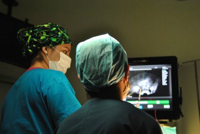
Minimally invasive Neurosurgery techniques:
NEUROSURGERY RESIDENCY
Nacional Medical Center "20 de Noviembre ISSSTE", México City Generation 2007
Title by Mexican Autonomous National University
Specialist License Number: 4991249
BOARD CERTIFIED, MEXICAL ASOCIATION OF NEUROLOGICAL SURGEONS Certificate Number: 811
ALLGEMEINES KRANKENHAUS – UNIVERSITÄTSKLINIKEN Vienna, Austria
Visiting M.D. in Functional Neurosurgery and Neuronavegation
JHO INSTITUTE OF MINIMALLY INVASIVE NEUROSURGERY Pittsburgh, USA
Fellow in Neuroendoscopy and Minimally Invasive Neurosurgery
MASTER IN NEUROSCIENCE AND BEHAVIOUR BIOLOGY University of Pablo de Olavide, Sevilla Spain
DIPLOMA IN HOSPITAL FINANCES AND ADMINISTRATION UNAM, Faculty of Superior Studies Campus Iztacala/p>
RESEARCH BACKGROUND
Experimental Microsurgery in animals
Institution: Proyecto Camina
Protocol: Spine regeneration with peripheral nerve or embryo spine trasplanstation in traumatic spine injury in rats
Date: August 1996 to June 1997
Social Service in Research
Institution: Proyecto Camina
Protocol: Spine regeneration with activated macrophagus trasplanstation in traumatic spine injury in rats
Date: August 1998 to June de 1999
Neurosurgery Final Tesis
Institution: ISSSTE
Clinical and Radiological Analysis of Pituitary Adenomas treated with Radiosurgery and
Fractionated Conformational Stereotactic Radiotherapy. Date: November 2006 to February 2007
TEACHING EXPERIENCE Lectures: Neuroanatomy fr Psycologist
School of Psycology. Anahuac University, Cancun. from August 2013 to October 2012
Lectures: Clinical Neurology, Neuroanatomy and Neurophysiology
Institution: Proyecto Camina, from August 1998 to June 1999
6. Seminarios /Congresos
Lectures
CNS Anatomy and Neuroblastoma
2th Medical Sanitary District, Cancun Qroo. 21 and 22 July 2011
Traumatic brain injury: Surgical treatment
I Medical Jornal 2009, HGR#17 IMSS Cancun Qroo. October 21 2009
Minimally Invasive Neurosurgery
III Academic Jornals, Quintaroo College of surgeons. Cancún Qroo. October 2nd 2009
Stereotactic and Neuronavegation "hand on workshops" (Organizer and Professor)
XX Mexican Neurological Surgeons Meeting, Galenia Hospital. Cancun Qroo. July 19 2009
Minimally Invasive Neurosurgery
Academic lectures, Galenia Hospital. Cancun Qroo. March 26 2009
Minimally Invasive Neurosurgery
Academic lecture. Amerimed Hospital. Cancún Qroo. November 26 2008
Brain Death Diagnosis
II Cancun Simposium "No step back" Organ Transplantation. Cancún Qroo. Oct 9 2008
Stereotaxy and Radiosurgery
Atizapan General Hospial, Mexico City. August 28 2008
Minimally Invasive Neurosurgery
Academic lecture, Cozumel Medical Center. Cozuel January 15 2008
Psichiatric Neurosurgery
Anahuac University, Cancun. November 22 2007
Minimally Invasive Neurosurgery
General meeting, General Hospital Cancun. October 23 2007
Minimally Invasive Neurosurgery
Academic lecture, Galenia Hospital. Cancun. October 10 2007
Neuroendoscopy
General meeting. General Hospital Cancun. July 20 2008
Neuroendoscopy
Neurosciences lectures. CMN 20 de Noviembre ISSSTE, Mexico City. January 3 2007
Clinical, Endocrinological and Radiological Analysis of Pituitary Adenomas treated with Radiosurgery and Fractionated Conformal Stereotactic Radiotherapy: Preliminary Report (presentation in English)
XXV International Course of Pituitary Adenomas, CMN 20 de Noviembre ISSSTE, Mexico
City. November 28 2006
Endoscopic Neurosurgery
XL Medical Meeting, Sociedad Médica Clínica Londres A.C. Mexico City. Nov. 24 2006
Adenomas de Hipófisis. Neurologic and Neurosurgical patient intensive course. CMN 20 de Noviembre, ISSSTE., Mexico City. October 24 2006
Minimally Invasive Neurosurgery
Ex-Nicolaitas Students Society, II Academic Meeting. Uruapan, Mich. October 12 2006
Arteriovenous Malformations and alternative treatments
Mexican Society of Neurological Surgery. Mexico City. March 7 2006
Neuromodulation
Mexican Society of Neurological Surgery. Mexico City. February 6 2006
Minimally Invasive Neurosurgery
La Sociedad de Ex alumnos Nicolaitas, 2da. Jornada Medica. Maravatio, Mich. July 5 2005
Advances in Radiosurgery
V Meeting of Neurocience Residents, CMN 20 de Noviembre , Mexico City. June 6 2004
Gamma Radiosurgery, 8 years in Mexico
IV Meeting of Neurocience Residents, CMN Siglo XXI, Mexico City. September 12 2003
Gamma Radiosurgery in Mexico
III Meeting of Neurocience Residents, CMN Siglo XXI, Mexico City. July 26 2002
Stereotactic surgical tecniques
V Academia week. Anahuac University, Mexico City. April 14 1999
Experimental paraplegia, experience in Proyecto Camina
III Academic week. Anahuac University, Mexico City. April 24 1997
Neurosurgical treatment of Epilepsy
I Academic week. Anahuac University, Mexico City. April 23 1995
Face Congenital Malformations
Academic meeting. Anahuac University, Mexico City. March 131994
MEETINGS AND COURSES
Eurospine 2011 Annual Meeting
Milan, Italy. October 19 to 21, 2011
International Course of Motion Preservation
Florence, Italy. October 17 to 18, 2011
XII National Meeting of AMCICO
AMCICO (Mexican Society of Spinal Surpeons)
Cancun Qroo. July 23 to 25, 2011
XXIV International Intradiscal Therapy Society (IITS)
Cancun Qroo. July 23 to 25, 2011
79th AANS Annual Meeting
Denver, Colorado. April 9 to 13, 2011
XXXIII Mexican Association of Neurology Annual Meeting
Cancún Qroo. November 15 to 21 2009
4th Annual Northwestern Radiosurgery Symposium
Northwestern Memorial Hospital, Chicago IL, USA. September 25 2009 (6.5 Credits)
XIV World Congress of Neurological Surgery
Boston MA, USA. August 30 to September 14 2009
XX Mexican Neurological Surgeons Meeting
Cancún Qroo, July 19 to 24 2009
Stereotactic and Neuronavegation "hand on workshops" (Organizer and Professor)
XX Mexican Neurological Surgeons Meeting, Galenia Hospital. Cancun Qroo. July 19 2009
Congress of Neurological Surgeons 58th Annual Meeting
Orlando FL, USA. September 20 to 25 2008 (24.50 credits)
Schloffer Conference and International Society of Pituitary Surgeons Meeting 2007
AKH Vienna Austria. September 7 to 10 2007
Master in Neurosciences and Behaviour Biology
Pablo de Olavide University, Sevilla Spain. January 2007 to October 2007
Member of "The Internacional Neuroendoscopy Study Group"
Pittsburg, EU. August 2006
Diploma in Hospital Finances and Administration
UNAM, Faculty of Superior Studies campus Iztacala. October 2005 to May 2006 (200 Hrs)
1st Minimally Invasive Neurosurgery International Meeting
Guadalajara General Hospital, April 24 to 29 2006 (20Hrs)
Workshop of Neuroendoscopy, Neurosonografy, Neuroendovascular Therapy
Guadalajara General Hospital, April 24 to 29 2006 (30Hrs)
I National Meeting of Neuroendoscopy
Mexican Society of Neurological Surgery. Mexico City. November 18 to 19 2005
European Workshop on Basic Techniques of Microsurgery and Cerebral
Revascularization
Medical University of Vienna, Austria. (AKH). October 21 to 26 2005
Selected topics in Neurocience
CMN 20 de Noviembre, ISSSTE. Mexico City. March 15 to 17 2005 (20 hrs)
Update in Gamma Knife
Médica Sur. Mexico City. November 11 to 14 2004 (15 hrs)
72nd ANNS Annual Meeting
Orlando Florida. May 1 to 6 2004 (21 credits)
XVII Mexican Meeting of Neurological Surgery
Monterrey N.L., July 19 to 25 2003
PUBLICATIONS
Maciel Rafael, Baltazar Jorge, Ramírez Vicente, González Armando y Cols. Técnicas Neuroendoscópicas: Indicaciones y procedimientos.
Revista de Especialidades Médico-Quirúrgicas Volumen 11, Num.3 Destrezas Clínicas. Pag. 63-67.
Maciel Rafael, Ramírez Vicente, Gonzalez Armando, Valdez Evangelina
Análisis Clínico, Endocrinológico y Radiológico de los Adenomas de Hipófisis Tratadas con Radiocirugía y Radioterapia Estereotáctica Conformacional Fraccionada: Estudio Preliminar.
Revista Mexicana de Neurociencias 2006: Volumen 7 Num. 2 Pag.573-580
Maciel Rafael, Baltazar Jorge, Garcia Silvia, Armando Gonzalez y cols.
Biopsia guiada por referencias anatómicas en una paciente con Leucoencefalopatía Multifocal
Progresiva (LMP).
Revista de Especialidades Médico-Quirúrgicas Volumen 10, Num.1 Destrezas Clínicas. Pag. 74-78.
7. Hospital(es) en donde atiende (destino Cancún, RM, Playa del Carmen)
PUBLIC PRACTICE
CANCUN GENERAL HOSPITAL "JESUS KUMATE RODRIGUEZ"
Cancun, Qroo.
PRIVATE PRACTICES
GALENIA HOSPITAL
Office 209, Cancún Quintana Roo
MÉDICA DEL CARMEN
Playa del Carmen Quintana Roo
COZUMEL MEDICAL CENTER (CMC)
Cozumel Quintana Roo.
RELEVANT PROCEDURES DONE IN CANCUN:
More than 600 neurosurgical procedures since I arrived to Cancun 6 years ago (May 14th, 2007).
40% of the cases are Brain, 60% spinal
problems.
PIONER IN THE SOUTH OF MEXICO, performing the First Neuronavegaction, stereotatic, neuroendoscopy, DBS surgery for Parkinson and distonía, Endoscopic pituitary adenoma resection in Cancun and the South of Mexico.
8. Acreditaciones y credenciales
MEMBER OF SOCIETIES AND GROUPS: Mexican Society of Neuroloical Surgery A.C.
Active Member
European Society for Stereotactic and Functional Neurosurgery (ESSFN)
Active Member
The Internacional Neuroendoscopy Study Group, Pittsburg, EU.
Active Member
European Association of Neurological Surgeons (EANS)
Active Member, Member Number 0570
American Association of Neurological Surgeon (AANS)
Active Member, October 2007
International Society of Pituitary Surgeons Meeting (ISPS)
Active Member
The Congress of Neurological Surgeons (CNS)
Active Member
NEUROSURGERY RESIDENCY
Nacional Medical Center "20 de Noviembre ISSSTE", México City
Generation 2007
Title by Mexican Autonomous National University
Specialist License Number: 4991249
BOARD CERTIFIED, MEXICAL ASOCIATION OF NEUROLOGICAL SURGEONS
Certificate Number: 811